
Table of Contents
Ultrasound Screening During Pregnancy | Trimester Scan Guide
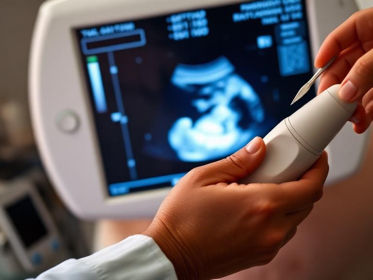
Routine Ultrasound Screening During Pregnancy is one of the most effective tools in modern obstetrics and fetal medicine. By converting high-frequency sound waves into real-time images, ultrasonography allows medical professionals to monitor embryonic development, evaluate fetal organ structures, assess placental blood supply, and confirm growth milestones without exposing the mother or fetus to ionizing radiation.
Over the past three decades, obstetric imaging has advanced from basic two-dimensional black-and-white structural checks to high-definition 3D/4D visual modeling, high-sensitivity Color Doppler hemodynamics, and specialized fetal echocardiography. Utilizing structured, trimester-specific ultrasound scans ensures potential issues—such as ectopic pregnancy, structural variations, placental insufficiency, or growth restriction—are identified early for optimal clinical management.

The Biological Principles & Safety of Diagnostic Ultrasound
Ultrasonography functions by emitting acoustic pulses—typically between 3.5 MHz and 12 MHz—from a piezoelectric transducer transducer probe. As sound waves travel through soft tissues, fluids, and bones, they bounce back as acoustic echoes. A computer processor calculates these echo intervals to build real-time cross-sectional images of the developing baby.
                  [ULTRASOUND SOUND WAVE TRANSMISSION MODEL]
                                       │
     ┌─────────────────────────────────┼─────────────────────────────────┐
     ▼                                 ▼                                 ▼
[Transducer Probe]            [Tissue Echo Reflection]        [Real-Time Rendering]
 Emits ultrasound pulses       Acoustic density changes        Generates cross-sectional
 through maternal abdomen       reflect sound back to probe     structural images on screen
Safety Record of Fetal Ultrasonography
- Zero Radiation Risk: Unlike X-rays or CT scans, ultrasound does not use ionizing radiation.
- ALARA Principle (As Low As Reasonably Achievable): Diagnostic ultrasound equipment is calibrated to minimize acoustic output energy and thermal index ($TI$), keeping power levels well below safe international threshold limits.
- Non-Invasive Nature: Standard transabdominal and transvaginal scans cause no pain or tissue injury to either the mother or developing baby.
Trimester-Wise Ultrasound Screening Roadmap
                    PREGNANCY ULTRASOUND SCANNING TIMELINE
                                       │
     ┌─────────────────────────────────┼─────────────────────────────────┐
     ▼                                 ▼                                 ▼
[FIRST TRIMESTER]             [SECOND TRIMESTER]                [THIRD TRIMESTER]
 (Weeks 6 to 13+6)             (Weeks 18 to 22)                  (Weeks 28 to 36+)
  • Early Viability Scan        • TIFFA Anomaly Scan              • Growth & Color Doppler
  • NT Chromosomal Scan         • Fetal Echocardiogram            • Biophysical Profile (BPP)
  • Crown-Rump Length (CRL)     • Cervical Length Check           • Amniotic Fluid Assessment
1. First Trimester Ultrasound Scans (Weeks 6 to 13+6)
Early pregnancy imaging establishes gestational age, verifies embryo viability, and provides early screening for chromosomal risk markers.
┌────────────────────────────────────────────────────────────────────────┐
│                   FIRST TRIMESTER ULTRASOUND SCANS                     │
├────────────────────────────────────────────────────────────────────────┤
│  1. Early Viability Scan (6–8 Weeks): Confirms intrauterine sac, heartbeat│
│  2. Nuchal Translucency (NT) Scan (11–13+6 Weeks): Checks neck fold thickness│
│  3. Nasal Bone Assessment: Evaluates markers for Trisomy 21 (Down syndrome)│
└────────────────────────────────────────────────────────────────────────┘
- Early Viability & Dating Scan (Weeks 6 to 8):
- Intrauterine Confirmation: Confirms the pregnancy sac is safely situated inside the uterine cavity, ruling out an ectopic pregnancy in the fallopian tubes.
- Cardiac Activity: Identifies the embryonic heartbeat, which typically ranges from 110 to 160 beats per minute (bpm).
- Crown-Rump Length (CRL): Measures the length of the embryo from the top of the head to the bottom of the torso, establishing an accurate Estimated Due Date (EDD) within a 3- to 5-day margin.
- Nuchal Translucency (NT) Screening Scan (Weeks 11 to 13+6):
- Translucency Measurement: Measures the fluid thickness under the skin at the back of the fetal neck. An increased NT thickness ($\ge 3.0 \text{ mm}$) may indicate an increased risk for Down syndrome (Trisomy 21), Trisomy 18, or congenital heart defects.
- Nasal Bone (NB) Check: Verifies the presence of the fetal nasal bone, an important marker used alongside double marker blood tests to refine chromosomal risk calculations.
2. Second Trimester Ultrasound Scans (Weeks 18 to 22)
Often termed the “Anomaly Scan,” this second-trimester check provides a comprehensive review of fetal anatomy and organ development.
┌────────────────────────────────────────────────────────────────────────┐
│                   SECOND TRIMESTER ULTRASOUND SCANS                    │
├────────────────────────────────────────────────────────────────────────┤
│  1. TIFFA Anomaly Scan (18–22 Weeks): Full organ anatomical evaluation │
│  2. Fetal Echocardiography: Detailed 4-chamber heart structural check   │
│  3. Transvaginal Cervical Length: Assesses risk for preterm delivery   │
└────────────────────────────────────────────────────────────────────────┘
- Targeted Imaging for Fetal Anomalies (TIFFA / Anomaly Scan):
- Neurosonography: Checks brain structures, including the lateral ventricles, cerebellum, and cisterna magna, to screen for conditions like spina bifida or hydrocephalus.
- Facial Profile: Examines the upper lip, nose, and eye orbits to rule out cleft lip and palate.
- Cardiovascular System: Evaluates the four heart chambers, along with the left and right ventricular outflow tracts.
- Gastrointestinal & Renal Systems: Confirms the presence of a filled stomach bubble, two functioning kidneys, and a normal urinary bladder.
- Musculoskeletal System: Verifies a complete 33-segment spine, normal curvature, and all upper and lower long bones.
- Placental Location: Locates the placenta relative to the internal cervical os to screen for placenta previa.
- Fetal Echocardiography (Weeks 20 to 24): A specialized ultrasound that evaluates cardiac blood flow, valve movement, and vessel connections in high-risk pregnancies (e.g., mothers with diabetes or family history of heart conditions).
3. Third Trimester Ultrasound Scans (Weeks 28 to Delivery)
Third-trimester scanning focuses on fetal growth velocity, placental adequacy, umbilical blood flow, and delivery readiness.
┌────────────────────────────────────────────────────────────────────────┐
│                   THIRD TRIMESTER ULTRASOUND SCANS                     │
├────────────────────────────────────────────────────────────────────────┤
│  1. Growth & Doppler Scan (28–34 Weeks): Measures weight & umbilical flow│
│  2. Biophysical Profile (BPP): Scores movement, tone, breathing, fluid │
│  3. Amniotic Fluid Index (AFI): Screens for Oligohydramnios / Polyhydramnios│
└────────────────────────────────────────────────────────────────────────┘
- Growth & Color Doppler Scan (Weeks 28 to 34):
- Biometric Measurements: Measures Head Circumference (HC), Biparietal Diameter (BPD), Abdominal Circumference (AC), and Femur Length (FL) to estimate total fetal weight via standard algorithms (e.g., Hadlock formula).
- Umbilical & Middle Cerebral Artery Doppler: Measures blood flow resistance ($S/D \text{ ratio}$, Pulsatility Index) to ensure adequate oxygen and nutrient delivery through the placenta.
- Biophysical Profile (BPP): Combines ultrasound observations with electronic fetal heart rate monitoring (Non-Stress Test), scoring four parameters on a 0 to 2 scale:
- Fetal Breathing Movements ($\ge 30 \text{ seconds}$ in 30 minutes)
- Gross Body Movements ($\ge 3 \text{ discrete body/limb movements}$)
- Fetal Muscle Tone ($\ge 1 \text{ episode of active extension and flexion}$)
- Amniotic Fluid Volume (Single deepest pocket $> 2 \text{ cm}$)
Anatomical Biometry Reference Parameters
Ultrasound equipment uses precise biometric measurements across gestation to calculate growth percentiles:
| Biometric Parameter | Abbreviation | Anatomical Reference Area | Clinical Significance |
| Crown-Rump Length | CRL | Top of head to bottom of breech | Most accurate dating parameter in early pregnancy (Weeks 6–13). |
| Biparietal Diameter | BPD | Transverse diameter of the fetal skull | Evaluates head growth and assists in gestational age calculation. |
| Head Circumference | HC | Outer perimeter of the skull | Evaluates cranial growth and neurological structure development. |
| Abdominal Circumference | AC | Outer perimeter of the fetal abdomen | Reflects liver size and subcutaneous fat stores; sensitive to intrauterine growth restriction (IUGR). |
| Femur Length | FL | Length of the femoral shaft bone | Assesses longitudinal skeletal development and limb proportion. |
| Amniotic Fluid Index | AFI | Sum of fluid depths in 4 quadrants | Normal range: $8 – 24 \text{ cm}$. Detects low fluid (oligohydramnios) or excess fluid (polyhydramnios). |
Patient Journey: Preparing for Your Pregnancy Ultrasound
1.1. Fluid Intake & Bladder Preparation:Pre-Exam Setup.
For early scans under 12 weeks, drink 3 to 4 glasses of water 1 hour prior to the exam without emptying your bladder. For later trimesters, normal hydration is sufficient.
2.2. Transducer Application & Acoustic Gel:Clinical Scanning.
Lie comfortably on the examination table. Water-based acoustic gel is applied to your abdomen to eliminate air gaps, allowing the transducer probe to transmit smooth sound waves.
3.3. Biometric Measurements & Anomaly Check:Real-Time Imaging.
The sonologist or fetal medicine specialist captures freeze-frame measurements, records fetal heart rates, evaluates internal organ structures, and assesses placental blood flow.
4.4. Image Documentation & Consultation:Reporting & Review.
You will receive printed ultrasound images and a structured report detailing gestational age, estimated fetal weight, placental grade, and anatomical findings for your doctor to review.
Comparative Analysis of Ultrasound Technologies
| Ultrasound Technology | Structural Resolution | Dimensionality | Primary Clinical Indication |
| 2D Gray Scale Imaging | High anatomical detail | 2D Cross-Sectional | Standard diagnostic scan for routine biometry, organ anatomy, and fluid checks. |
| 3D Static Ultrasound | High surface detail | 3D Volumetric | Provides detailed views of facial features, palate structure, and skeletal variations. |
| 4D Real-Time Motion | Continuous dynamic view | 3D + Time | Evaluates fetal movement, facial expressions, and complex behavioral patterns. |
| Spectral Color Doppler | Dynamic vascular flow | Velocity Waveforms | Evaluates placental sufficiency, umbilical artery flow, and fetal hemodynamic adaptation. |
| Transvaginal (TVS) | Ultra-high close range | 2D High-Frequency | Ideal for early viability checks, ectopic pregnancy screening, and precise cervical length measurements. |
Estimated Cost Guidelines for Pregnancy Ultrasound Scans in India
Ultrasound pricing varies depending on the technology used, diagnostic center accreditation, and fetal medicine specialization:
| Ultrasound Screening Category | Estimated Cost Range (INR) | Key Details & Deliverables |
| Early Viability / Dating Scan | ₹1,000 – ₹2,000 | Confirms heartbeat, gestational sac, and Crown-Rump Length (CRL). |
| Nuchal Translucency (NT) Scan | ₹2,000 – ₹3,500 | Includes NT fluid measurement, nasal bone check, and soft marker assessment. |
| TIFFA Anomaly Scan (2D/3D) | ₹2,500 – ₹4,500 | Comprehensive multi-organ anatomical review and placental evaluation. |
| Growth & Color Doppler Scan | ₹2,500 – ₹5,000 | Fetal weight calculation, amniotic fluid index (AFI), and vascular flow studies. |
| Fetal Echocardiogram | ₹3,000 – ₹6,000 | Specialized pediatric cardiac evaluation of four chambers and outflow tracts. |
| Full 9-Month Ultrasound Package | ₹8,000 – ₹16,000 | Comprehensive package covering all four standard trimester scans. |

Frequently Asked Questions (FAQs)
1. Why is ultrasound screening during pregnancy necessary?
Ultrasound screening provides essential diagnostic information by confirming gestational age, checking fetal cardiac activity, detecting structural anomalies, evaluating placental location, and monitoring fetal growth and blood flow.
2. How many routine ultrasound scans are required during a low-risk pregnancy?
A standard low-risk pregnancy includes 4 essential scans: Early Viability Scan (6-8 weeks), NT Scan (11-13+6 weeks), Anomaly/TIFFA Scan (18-22 weeks), and Growth Doppler Scan (28-34 weeks).
3. Is ultrasound screening safe for the baby and mother?
Yes. Ultrasound uses high-frequency sound waves rather than ionizing radiation (X-rays). Decades of clinical evidence demonstrate that medical ultrasound is completely safe for both mother and fetus.
4. What is the Nuchal Translucency (NT) Scan and when is it performed?
The NT scan is performed between 11 weeks and 13 weeks + 6 days to measure the fluid-filled space at the back of the baby’s neck, evaluating risk for chromosomal conditions like Down syndrome.
5. What is a TIFFA or Anomaly Scan?
The TIFFA (Targeted Imaging for Fetal Anomalies) scan, done between 18 and 22 weeks, is a detailed anatomical evaluation that examines fetal organs, brain structures, heart chambers, spine, kidneys, facial features, and placental positioning.
6. Do I need a full bladder for every pregnancy ultrasound?
A full bladder is generally required for transabdominal scans in the first trimester (under 12 weeks) to lift the uterus into view. In later trimesters, amniotic fluid provides a clear acoustic window, so a full bladder is usually unnecessary.
7. What is the difference between Transabdominal (TAS) and Transvaginal (TVS) Ultrasound?
Transabdominal ultrasound scans through the lower abdomen with gel. Transvaginal ultrasound uses a small internal probe to provide higher-resolution imaging during very early pregnancy or to measure cervical length.
8. What is a Color Doppler Ultrasound during pregnancy?
A Color Doppler scan measures blood flow velocity through the umbilical artery, uterine arteries, and fetal middle cerebral artery, evaluating placental efficiency and oxygen delivery.
9. How much does an ultrasound scan cost during pregnancy in India?
In India, basic viability scans range from ₹1,000 to ₹2,000. Specialized NT and TIFFA anomaly scans range from ₹2,000 to ₹4,500, while advanced Doppler growth scans cost between ₹2,500 and ₹5,000.
10. What is 3D/4D ultrasound imaging?
3D ultrasound renders three-dimensional static images of the fetus, while 4D ultrasound adds real-time motion. These scans assist in evaluating facial or skeletal structures and provide clear visual views.
11. Can an ultrasound scan detect all birth defects?
While advanced TIFFA scans detect approximately 70% to 85% of major structural anomalies, some microscopic, metabolic, functional, or late-developing conditions cannot be detected via ultrasound alone.
12. Why is cervical length measured during a pregnancy ultrasound?
Measuring cervical length helps evaluate the risk of preterm labor. A shortened cervix (under 25 mm) in the second trimester may require clinical intervention, such as progesterone support or cervical cerclage.
13. What is a Biophysical Profile (BPP) ultrasound test?
A Biophysical Profile combines a Non-Stress Test (NST) with ultrasound scoring of fetal breathing movements, body tone, gross limb movement, and amniotic fluid volume to evaluate overall fetal well-being in the third trimester.
14. What does placenta previa mean on an ultrasound report?
Placenta previa indicates that the placenta covers or lies close to the internal opening of the cervix. It requires careful monitoring in the third trimester to determine whether a safe vaginal delivery is possible.
15. How long does a routine pregnancy ultrasound take?
A routine viability or growth scan takes about 15 to 20 minutes. Complex screening scans like the TIFFA anomaly scan or detailed fetal echocardiography take 30 to 45 minutes.


Leave a Reply